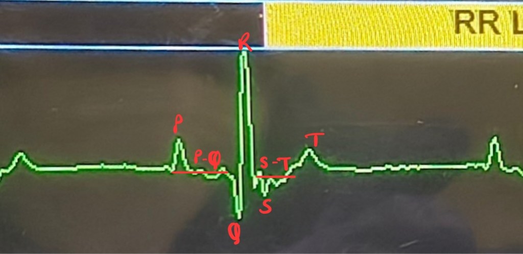
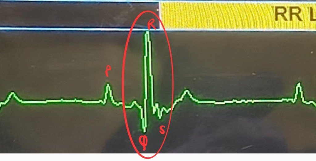
Electrocardiogram has two components, a baseline or isoelectric line which represents pause between two heartbeats or absence of electrical activity, another component are positive and negative deflections or waves formed by depolarization (contraction) and relaxation (repolarization) of heart chambers1.
Mammalian heart has two small chambers “atria” at the top and two larger chambers “ventricles” at the bottom2.
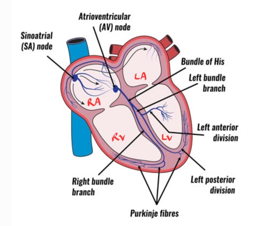
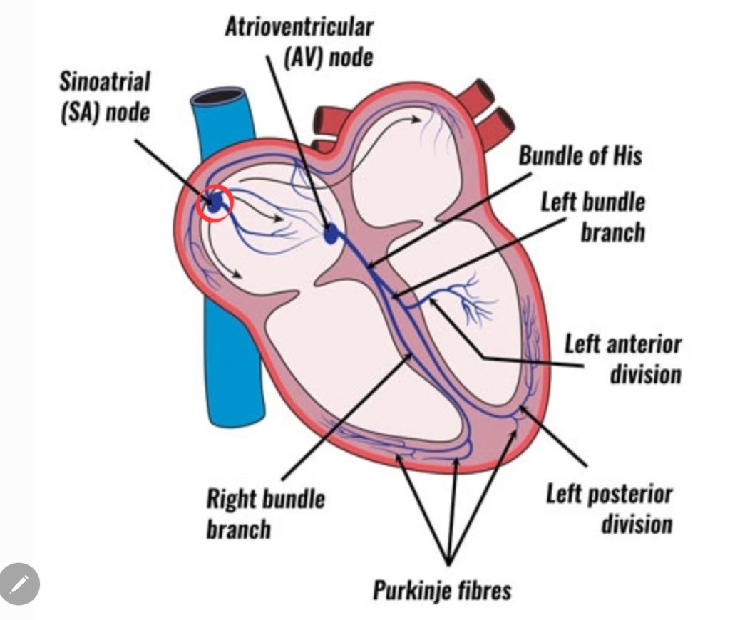
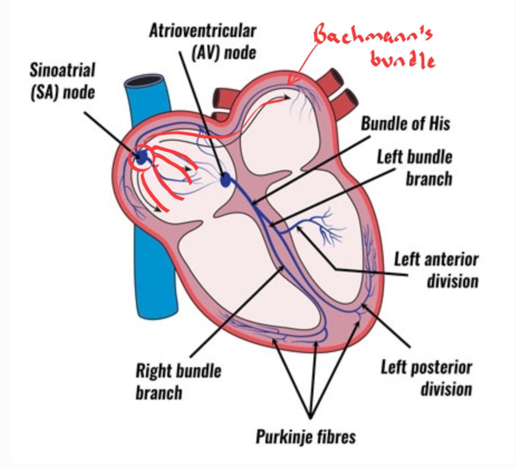
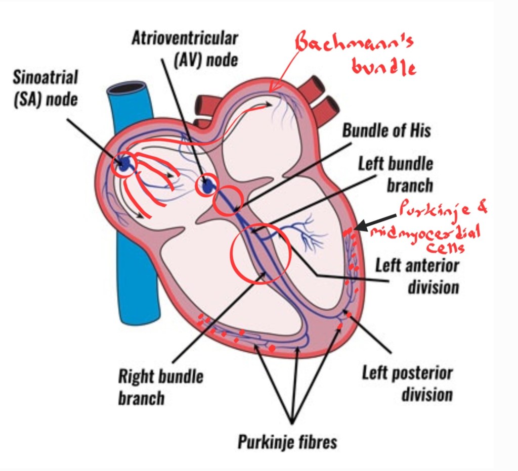
Sinoatrial or SA node is a small group of specialized right atrial muscle cells which is the dominant pacemaker of the heart. This node generates the primary electric impulse. This event does not show up on ECG since few cells are involved and the electromotive force is not large enough to be recorded by ECG leads1.
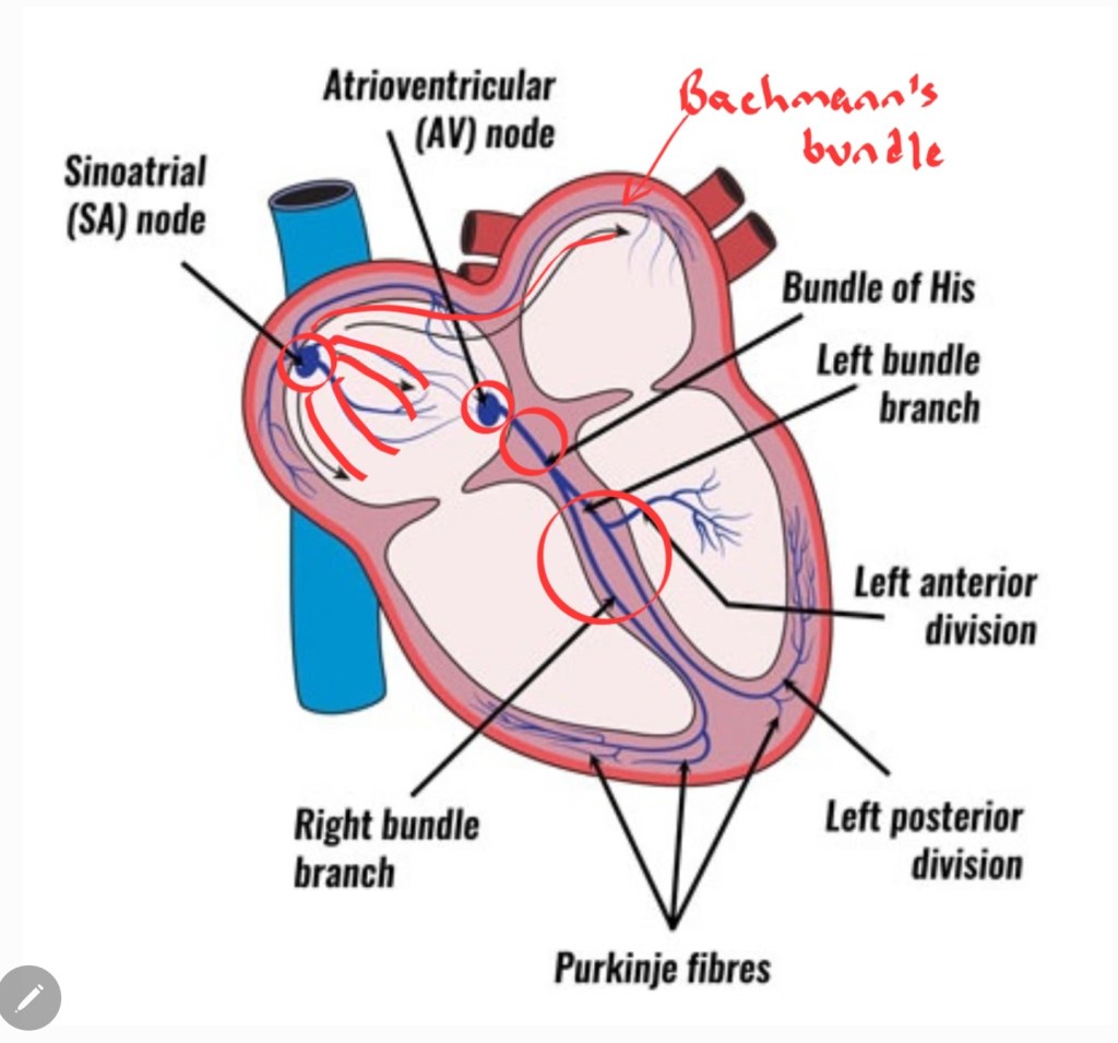
When SA node activates, a wave of depolarization passes throughout right atrium and it goes to left atrium by this specialized conducting pathway, called Bachmann’s bundle2.
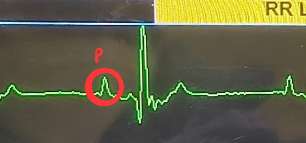
Depolarized atrial cells form a positive deflection, the first wave of a heartbeat, called P wave. In adult giant breed dogs you may see a bifid P wave which shows individual depolarization of right and left atrium1.
This electric impulse reaches Atrioventricular or AV node, which is a group of specialized cells to conduct electric impulse in interatrial septum, the wave of depolarization passes along the bundle of His and the right and left bundle branches till Purkinje fibers1.
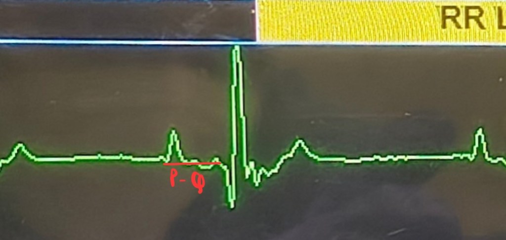
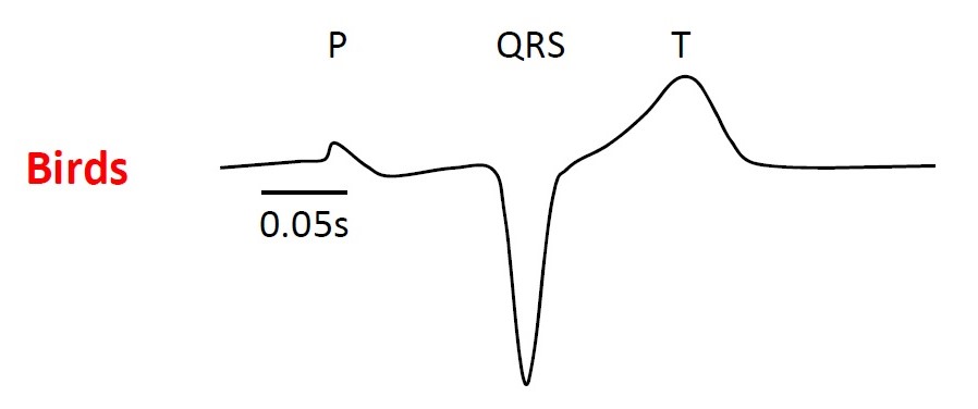
The time it takes for this propagation from atria to ventricles is shown as PQ segment. It begins from the onset of P wave to the onset of QRS complex1. During this time both atria contract and empty blood into respective ventricles.
Purkinje network spreads this impulse from the apex to the ventricular cells and depolarizes the whole ventricular muscle mass. This depolarization forms the QRS complex1.
Purkinje cells and midmyocardial cells at the end of Purkinje fibers prevent recycling and reentry of impulse in ventricular muscle2.
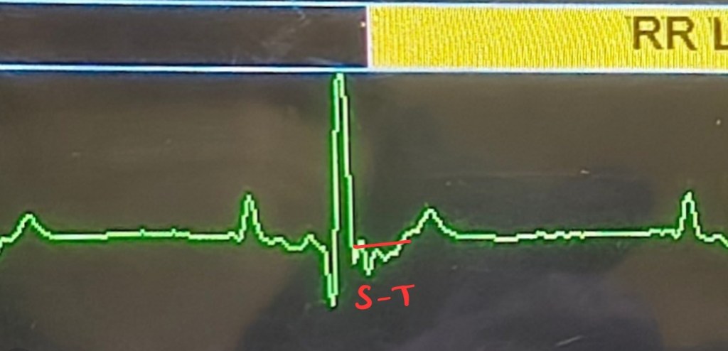
The line between the negative S deflection and positive T wave is ST segment, when both ventricles contract and push blood out of the chambers. From right ventricle blood goes to the lungs through pulmonary arteries and from left ventricle it goes to aorta2.
Phase of repolarization follows, where ventricles relax from a contracted state and form a positive deflection, T wave. Repolarization of atria is represented as Ta wave but it is overlapped by the large QRS complex1. This is when atria fill with blood, right atrium by superior and inferior vena cava and left atrium gets blood from lungs through pulmonary veins2. And the cycle continues.
Connect with me on LinkedIn and Instagram. Subscribe to the AnesWise blog newsletter for future content notifications. Kindly leave a feedback as it helps improve the blog. Feel free to ask your doubts in the comment section or email it to dr.sahilmehta@outlook.com
Edited by Prajakta Alase
Citations:
- Bartholomew, K.J., 2024. Electrocardiography. Veterinary Anesthesia and Analgesia: The Sixth Edition of Lumb and Jones, pp.187-196.
- Muir, W.W., 2024. Cardiovascular physiology. Veterinary anesthesia and analgesia: the sixth edition of Lumb and Jones, pp.615-666.
Image source: https://www.teachpe.com/anatomy-physiology/the-heart-conduction-system


Leave a comment