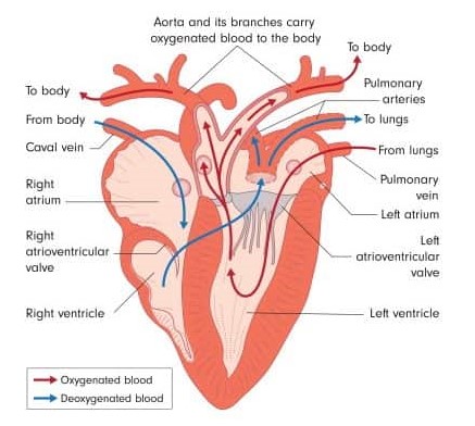
Anatomy and electrophysiology of mammalian and avian heart have a few similarities and many differences. Avian heart is four chambered, but relative to body mass, it is larger in size, has higher stroke volume, cardiac output and blood pressure, when compared with mammals.
Conduction system consists of SA node, AV node and Purkinje fibers, with a major difference between mammals and birds that, Purkinje fibers penetrate ventricles completely from endocardium to epicardium which helps with synchronous ventricular contraction at high heart rates. This modification is evident on avian ECG.
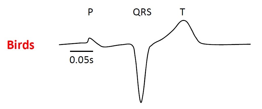
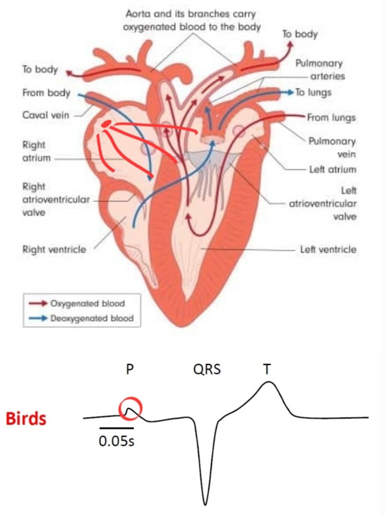
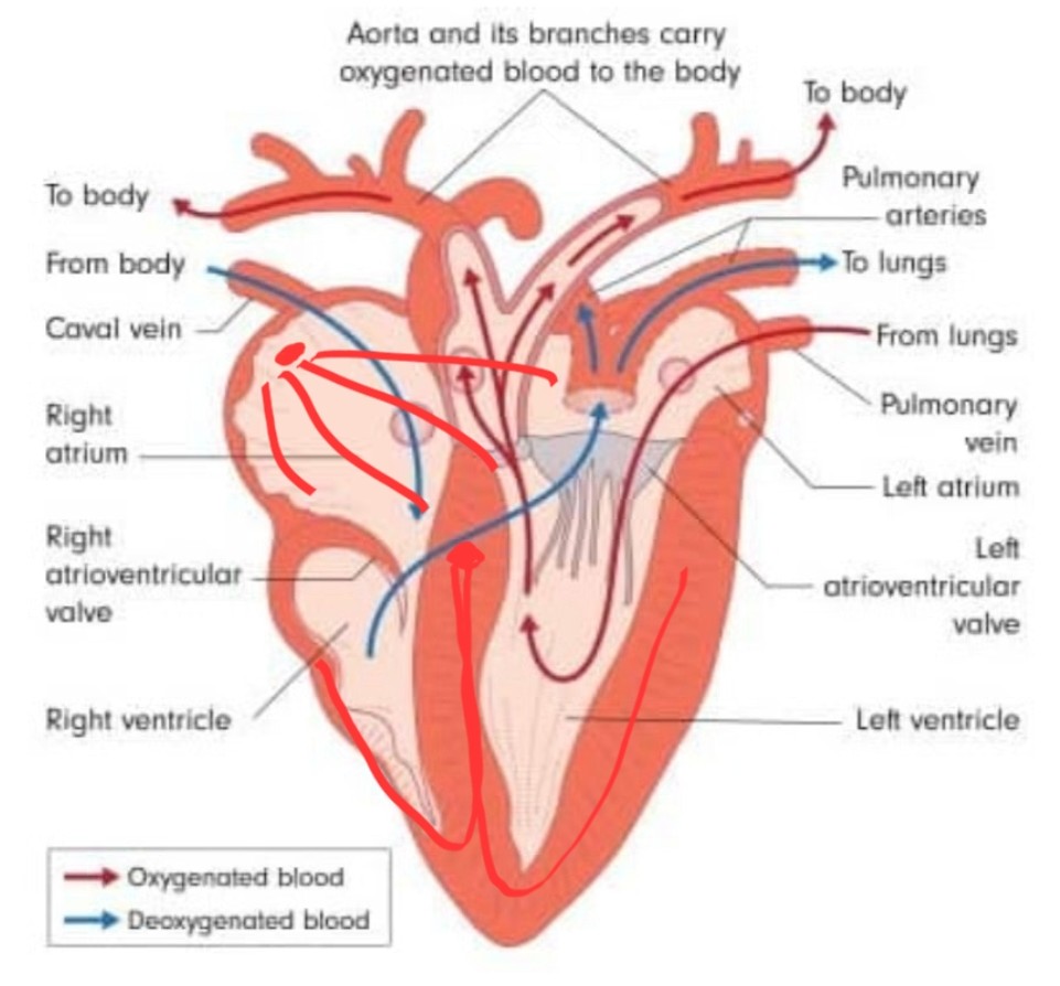
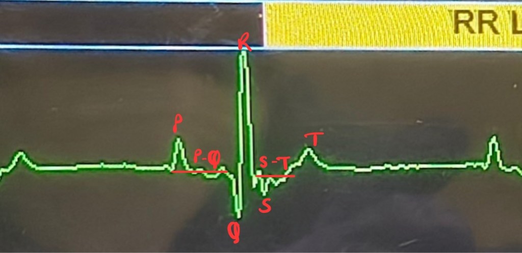
It begins with impulse generated by SA node in right atrium, traverses from right to left and towards cardiac apex, which depolarizes both atria and gives a positive deflection- P wave on ECG.
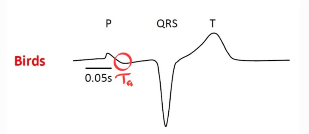
Right after P wave, a negative deflection is seen which is Ta wave. It represents repolarization of atria.
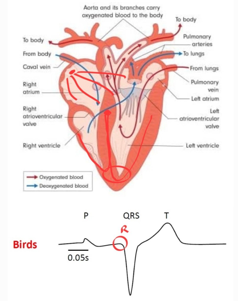
Impulse reaches AV node and spreads across ventricular myocardium via Purkinje fibers.
Ventricles first depolarize at the level of apex which is another small positive deflection- R wave.
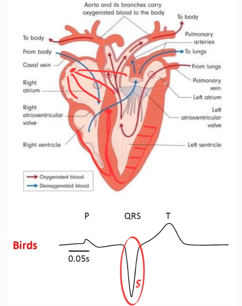
Purkinje network penetrates deep into the myocardium in birds, which carries impulse from myocardium towards endocardial surface. That is in contrast to mammalian heart, where impulse goes through endocardial surface to epicardial side. This is the reason for large and inverted QRS/rS complex on avian ECG. The deepest wave is S wave.
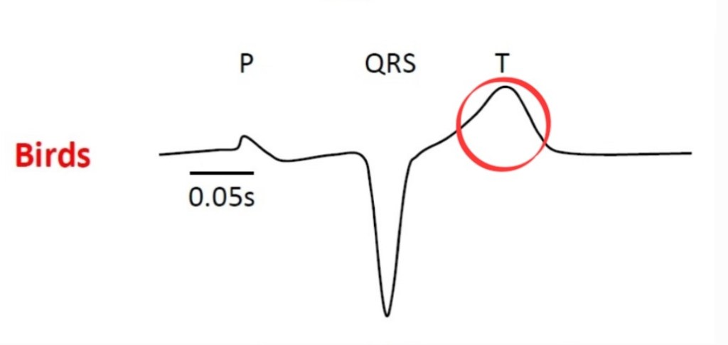
ST segment is either very short or absent. Few bird species show ST slurring where T wave merges with S wave at baseline. Ventricular repolarization is a positive deflection- T wave.
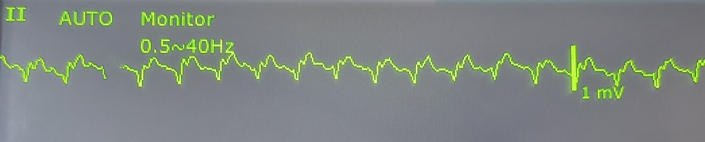
On standard multipara monitors, avian ECG waves appear very small and indiscernible due to high heart rate. Ideal speed to observe/record ECG of birds is 100-200mm/sec, instead of 50mm/sec used for small animals. Despite these limitations, knowing normal bird ECG helps a lot with monitoring cardiac rhythm under anesthesia. Developing field of exotic species cardiology has found species differences in ECG which helps determine whether certain ECG is normal for that bird.
Few species have T wave right at the base of S wave. It is called S-T slurring, commonly seen in pigeons, parrots and parakeets.

P-on-T wave phenomenon, where P wave is superimposed on T wave is considered normal in Amazon parrots and few gray parrots.
There are many cardiac arrhythmias diagnosed in various bird species which can be used to assess anesthetic risk.
Connect with me on LinkedIn and Instagram. Subscribe to the AnesWise blog newsletter for future content notifications. Kindly leave a feedback as it helps improve the blog. Feel free to ask your doubts in the comment section or email it to dr.sahilmehta@outlook.com
Edited by Prajakta Alase
Citations
- Reddy, B.S. and Sivajothi, S., 2017. Avian electrocardiography a simple diagnostic tool. International Journal of Avian & Wildlife Biology, 2(2).
- Zandvliet, M.M., 2005, January. Electrocardiography in psittacine birds and ferrets. In Seminars in avian and exotic pet medicine (Vol. 14, No. 1, pp. 34-51). WB Saunders.
- https://study.com/academy/lesson/bird-circulatory-system-function-structure.html
Image credits:
- Boukens, B.J., Kristensen, D.L., Filogonio, R., Carreira, L.B., Sartori, M.R., Abe, A.S., Currie, S., Joyce, W., Conner, J., Opthof, T. and Crossley II, D.A., 2019. The electrocardiogram of vertebrates: Evolutionary changes from ectothermy to endothermy. Progress in biophysics and molecular biology, 144, pp.16-29.
- https://earthlife.net/the-bird-circulatory-system-heart-blood/


Leave a comment